abspathology
Antonio Schütz A service provide by abspathologie ++ Die Pathologie dass der Probleme hat, ist für Sie zu sehen ++ abspathology ++ A service provide by abspathology ++ abspathogie ++ The pathology that present problem is presented to you ++ abspatholgy ++ A service provide by abspathologie ++ abspathology ++ A mamata que voce somente ouviu falar é explicada para você++ abspathology++ A service provide by abspathologie ++ abspathology ++ Die Pathologie dass der Probleme hat, ist für Sie zu sehen ++ abspathology ++ A service provide by abspathology ++ abspathologie++The pathology that present problem is presented to you ++ abspathology ++ A service provide by abspathologie ++ abspathology ++ A mamata que voce somente ouviu falar é explicada para você++ abspathology ++ A service provide by abspathology ++ abspathologie ++ Die Pathologie dass der Probleme hat, ist für Sie zu sehen ++ abspathology ++ A service provide by abspathologie ++ The pathology that present problem is presented to you ++ abspathologie ++ A service provide by abspathology ++ abspathologie ++ A mamata que voce somente ouviu falar é explicada para você++ abspathologie ++ A service provide by abspathology ++ abspathologie ++ Die Pathologie dass der Probleme hat, ist für Sie zu sehen ++ abspathology ++ A service provide by abspathologie ++ The pathology that present problem is presented to you ++ abspathologie ++ A service provide by abspathology ++ abspathologie ++ A mamata que voce somente ouviu falar é explicada para você++ abspathology ++ A service provide by abspathology ++ abspathology ++ Die Pathologie dass der Probleme hat, ist für Sie zu sehen ++ abspathology ++ A service provide by abspathologie ++ abspathology ++ The pathology that present problem is presented to you ++abspathologie ++ A service provide by abspathology ++ A mamata que voce somente ouviu falar é explicada para você++ Principal themes
▼ abs pathology Letters Report of case Article Brazilian academic theses
Histhological Malignancy System: WHO x MGSH (criticism)
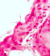
In Portuguese Stage of invasion grade 3
PARA ABS PATHOLOGY: O trabalho do Dr. Schütz avançou na compreensão desta controvertida área do conhecimento e, ainda, escassamente estudada. Entretanto, confesso que ao ler este artigo tive dificuldade em compreender como poderia a graduação histológica de malignidade fornecer uma previsão sobre o prognóstico e a tendência para a disseminação metastática, considerando que até a presente data não há um marcador biológico ou histopatológico desta tendência, ou que viabilize essa previsão. Por que foram excluídos os casos em que a lesão acometeu mais de uma região anatômica? Tendo em vista este critério de seleção, não haveria comprometimento da representatividade da amostra estudada? Além disso, a metodologia empregada, também, não menciona se foi utilizada ou não a "calibração" dos examinadores e/ou estudo de reprodutibilidade dos diferentes parâmetros histológicos avaliados. Também não compreendi com base em que o autor concluiu que o sistema histológico de graduação de malignidade do tipo multifatorial teria maior precisão do que o sistema monofatorial, baseado apenas na diferenciação celular da neoplasia. Além disso, pode ter ocorrido erro avaliativo na análise do parâmetro histológico profundidade de invasão, pois tal como foi salientado por Larsen e colaboradora (1978) [1], em relação ao carcinoma de laringe, muitos casos de biópsias incisionais são superficiais, não possibilitando a distinção entre os graus três e quatro desse parâmetro; por conseguinte, causando decréscimo na graduação histológica de malignidade do carcinoma, bem como interferindo na previsão de metástases para os linfonodos cervicais e prognóstico. Portanto, não seria mais racional omitir-se a avaliação desse parâmetro por se tratar de biópsia incisional, tal como é rotineiramente praticado nas teses defendidas no âmbito da Faculdade de Odontologia da Universidade de São Paulo (FO/USP)? REFERENCE [1] Larsen KH, Graem N, Larsen KIM, Larsen UM Clinical Relevance of Histological Grading of Cancer of the Larynx. Acta Path. Microbial. Scand Sect A 86: 499, 1978. Réplica de abspathology: Se bem compreendi os questionamentos do professor de Natal, Rio Grande do Norte, Brasil, eles foram, inicialmente, dirigidos à compreensão da proposição de minha tese de mestrado. A despeito professor brazileiro estar, parcialmente, correto na afirmação de que ainda não há um marcador biológico específico, ou qualquer variável histológica que possibilite, com certeza, antever o aparecimento de metástases, bem como fazer uma previsão sobre o prognóstico do paciente portador de carcinoma oral vários trabalhos publicados desde 1978 reportam uma correlação estatística positiva entre a graduação histológica de malignidade e a freqüência de metástases, e negativa para a sobrevida. Além do que, algumas dessas variáveis histopatológicas têm sido relacionadas com a invasividade do tumor aos tecidos normais do paciente. Dentre elas o modo de invasão, a profundidade de invasão, a invasão vascular, a embolização vascular e a resposta imune do paciente foram reportadas como as de maior valor para o prognóstico e previsão sobre a disseminação das metástases. Como a proposição de nosso estudo foi estudar a graduação histológica de malignidade, foi utilizada uma amostragem estratificada, segundo o grau histológico. Tal amostragem é indicada quando se trabalha com amostras provenientes de um grupo amostral restrito (pacientes atendidos no Departamento de Patologia e Diagnóstico bucal da Faculdade de Odontologia da Universidade Federal do Rio de Janeiro - FOUFRJ); portanto, perfeitamente representativa da população estudada. O critério da exclusão de lesões que se localizavam em mais de uma região anatômica, sem origem definida, ao contrário do que questiona o professor de Natal - tornou mais acurada a identificação da localização anatômica de origem do carcinoma oral. A diferença não significativa entre as lesões de assoalho de boca (17 casos) e língua (15 casos), verificada pelo nosso estudo, apontando-as como as regiões anatômicas preferencialmente acometidas pelo carcinoma oral, também, foi corroborada por dados recentes, divulgados pelo INCA/RJ (Instituto Nacional do Câncer, RJ) e pelo professor da UFRJ, Abel Silveira Cardoso e colaboradores (Sobe-2002, PR, Brasil), no âmbito da Faculdade de Odontologia da UFRJ, que as apontam, outrossim, como as mais freqüentes regiões anatômicas de acometidas pelo carcinoma oral no Estado do Rio de Janeiro, Brasil e FOUFRJ, respectivamente. O questionamento referente ao estudo da reprodutibilidade das diferentes variáveis estudadas é procedente, contudo, como o nosso trabalho foi direcionado para a graduação histológica e não para a análise de cada parâmetro, individualmente, ela não foi realizada. Indubitavelmente, tal estudo teria sido importante para a confirmação da análise microscópica descritiva por nós realizada. Quanto à "calibração" dos examinadores, nós entendemos que ela seria contra-indicada, visto que esse tipo de estudo requer precisamente a não comunicação entre os examinadores tal como foi feita pela família Larsen e colaboradora (1978). Além disso, os critérios histopatológicos do sistema de graduação de malignidade, por nós desenvolvidos, com base no sistema de Anneroth e colaboradores (1987), eram suficientes para uma análise acurada e calibrada. Além disso, estudos anteriores comprovaram que o sistema de Anneroth e cols. (1987) é reprodutível, particularmente, quando usado conjuntamente com o corte seriado ou semi-seriado, tal como foi feito em nosso estudo. Nós entendemos que os sistemas de graduação de malignidade, baseados, tão-somente, na população celular do carcinoma, tal como a classificação da Organização Mundial de Saúde (WHO), estão sujeitos a erro avaliativo, pois o percentual de células maturas (ceratinizadas) ou apresentando polimorfismo nuclear e/ou celular, bem como o número de mitoses varia de campo para campo, nas diferentes profundidades do carcinoma, examinado ao microscópico. Tal dificuldade de avaliação não é observada quando analisamos os parâmetros modelo e estágio de invasão, em pequeno aumento (x 5), que possibilita, no corte semi-seriado ou seriado, a identificação da região de maior invasão. O estudo da variável infiltrado inflamatório ou resposta imune, também, com o emprego de técnicas de coloração especial (imunohistoquímica), conjuntamente com a utilização desses cortes histológicos, é um outro importante parâmetro para o estudo da competência imunológica do paciente à neoplasia. Em parte, a família Larsen e colaboradora (1978) tinham razão em afirmar que, em determinados casos, por serem superficiais, as biópsias incisionais não propiciam a distinção entre os graus 3 e 4 da variável estágio de invasão. Entretanto, a extrapolação feita pelo professor doutorando do curso de Patologia Bucal da Universidade Federal do Rio Grande do Norte, Natal, Brasil, que em decorrência disso ocorreria um decréscimo na graduação histológica de malignidade dos carcinomas, de modo a interferir na previsão de metástases para os linfonodos regionais e prognóstico, não estão corretas. Senão vejamos com o seguinte cálculo matemático: considerando que eram 6 parâmetros analisados, graduados de 1 a 4 pontos, totalizando 24 pontos, com escore histológico total variando de 1 a 4 pontos (de 6-24/6). A atribuição do grau 4 para todos os 6 parâmetros histopatológicos implicaria em um escore histológico de 4 pontos (escore real). Admitindo que o parâmetro estágio de invasão (escore real 4) tenha sido erroneamente graduado, para menos, com os graus 2 (-0.32 pontos) ou 3 (-0.16 pontos), o escore histológico final seria 3,68 e 3,84, respectivamente. Ainda bastante superior aos 2,5 pontos necessários para classificá-los como de alto escore histológico de malignidade. Portanto, indicativo de sua natureza agressiva, mal prognóstico e alta probabilidade de disseminação metastática; conservando, portanto, o valor prognóstico desse parâmetro, tal como reportado por Anneroth et al. (1987). Poderia o ilustre e erudito professor pós-graduando do curso em Patologia Bucal da Universidade Federal do Rio Grande do Norte, Natal, Brasil, contra-argumentar, que esse cálculo somente seria válido para os graus extremos, nos quais, todos os parâmetros histopatológicos apresentariam o grau 4. Novamente, discordaria do erudito professor. Para os casos em que os parâmetros histopatológicos apresentassem grau 3 e o parâmetro estágio de invasão apresentasse grau 4, o escore histológico real seria 3.16 (3 x 5 +4). Caso o parâmetro estágio de invasão tivesse sido graduado para menos com os graus 3 (-0.16) ou 2 (.0.32), o escore final seria 3 e 2.84 pontos, respectivamente, ainda superior aos 2.5 pontos necessários para classificá-los como de alta graduação histológica de malignidade. Não concordando com esse cálculo, o ilustre professor poderia replicar, como fez, erroneamente, na reunião da SOBE (2002), que o valor na diferença de 1 grau com relação ao parâmetro estágio (profundidade) de invasão seria 0.25 (1/4) e não 0.16666 ou 0.17 como mencionei. Como fiz na SOBE (2002), novamente, e pela terceira vez, discordaria do sapiente professor, uma vez que a diferença de 1 grau no escore histológico é 0.166 ou 0.17 (1/6). Mesmo assim, faremos o cálculo baseado nesse raciocínio errôneo, imaginado pelo caro professor de Natal, RN, Brasil. Considerando 5 (número de parâmetros analisados) x 3 (grau atribuído a cada parâmetro) = 15 + 4 (grau atribuído ao parâmetro profundidade de invasão) = 19/6 = 3,16 (escore histológico real). Desse valor bastaria diminuir 0.25 ou 0.50, atribuído à graduação para menos (graus 3 e 2) do parâmetro profundidade de invasão (3,16 - 0.25 e 0.50), e teríamos os escores histológicos de 2,91 e 2,66, respectivamente. Portanto, novamente repito: "ainda, superior aos 2.5 pontos necessários para considerá-los como de alta graduação histológica de malignidade (grau III), e, portanto, indicativa de sua natureza agressiva, mal prognóstico e alta probabilidade de disseminação metastática, conservando o valor prognóstico desse parâmetro (escore histológico), tal como reportado por Anneroth et al. (1987)". Para justificar a minha afirmação: "de que a graduação histológica inferior a real implica tão somente na diminuição no escore histológico de 0.16666 pontos, e não 0.25 pontos como afirmou o Professor doutor da UFRN"; faço um novo cálculo matemático: com o grau 3 atribuído ao parâmetro profundidade de invasão, o escore histológico seria (5 x 3 + 3) = 18/6 = 3 e o escore real seria (5x3+4)=19/6 = 3.1666. Portanto, a diferença de 1 grau para cada parâmetro implica em um valor de somente + ou - 0.1666 ou 0.17 como afirmei, não comprometendo o valor prognóstico do escore ou do grau histológico de malignidade dos carcinomas orais, tal como afirmaram a família Larsen e colaboradora, corroborado pelo professor de Natal, RN, Brasil, bem como tem sido reportado em hipóteses defendidas na FO/USP. Além disso, a variável estágio de invasão é perfeitamente identificável, segundo os parâmetros histopatológicos de Anneroth e Hansen (1984) [2], bastando para isso, tão-somente, identificar (grau 3) ou não (grau 4) a presença da lâmina própria; ou a presença das células neoplásicas nas proximidades de glândulas, músculo ou tecido adiposo (grau 3). Particularmente não concordo com a exclusão de qualquer um dos parâmetros histopatológicos reportados por Anneröth e colaboradores (1987) [3], tal como foi sugerido e empregado em hipóteses defendidas no âmbito da Faculdade de Odontologia da Universidade de São Paulo (FO/USP) - mesmo em se tratando de biópsias incisionais. Contudo, reconheço que se assim se proceder, tão-somente, nos casos em que as diferenças no estágio de invasão entre os graus 3 e 4 não puderem ser estabelecidas, tal omissão (escore 4) ou a graduação para menos (escore 3,83), não interferirá na gradação histológica de malignidade do carcinoma, pois não é suficiente para os remover da classificação de tumores de alto escore ou graduação histopatológica de malignidade (graus 3 ou 4), tal como re portado na revisão de Anneroth et al (1987), cujo valor limítrofe é 2,5; mesmo, utilizando-se qualquer um dos escores histológicos (4 ou 3,83) ou critérios de avaliação mencionados, ou seja, exclusão ou graduação para menos. Contudo, considerando que, três dos quatro graus do parâmetro estágios de invasão são identificáveis, mesmo em biópsias incisionais, discordo da exclusão da análise do parâmetro estágio de invasão, quando a lesão se apresenta com esses graus, bem como da omissão da avaliação do parâmetro resposta linfo-plasmocitária, (que tem sido omitida em sucessivas hipóteses defendidas na Faculdade de Odontologia da Universidade de São Paulo) FOUSP, visto que, quando acentuada, possivelmente, é indicativa da resposta imune do paciente à neoplasia. Também não concordo com a não avaliação do escore histológico, reportado em algumas outras hipóteses defendidas no âmbito dessa mesma Faculdade, pois, particularmente, nos casos em que a maioria dos parâmetros histopatológicos se apresenta com diferentes graus, o escore histológico é decisivo no estabelecimento do grau histológico de malignidade do carcinoma. REFERÊNCIAS [1] Larsen KH, Graem N, Larsen KIM, Larsen UM Clinical Relevance of Histological Grading of Cancer of the Larynx. Acta Path. Microbial. Scand Sect A 86: 499, 1978. [2]Anneroth G, Hansen LS A methodologic study of histologic classification and grading of malignancy in oral squamous cell carcinoma. Scand. J. Dent Res. 92 : 448, 1984. [3] Anneroth G, et al Review of the literature and recommended system of malignancy grading in oral squamous cell carcinoma. Scand J. Dent Res 95: 229, 1987
In English Stage of invasion grade 4
For ABS pathology: The work of Dr. Schütz advanced in the comprehension of this controversy field of the knowledge and scarcely studied. However, I confess that at the to read this work had difficulty for understanding as would be able to histological gradation of malignance supply a forecast about the prognostic and tendency to dissemination of metastases, since presently there is not any biological marker or histological of this tendency, or that makes feasible this forecast. Why were excluded the cases in that the neoplasm attacked more of an anatomical region? Having in mind this criterion of selection, there would not be a decrease of representation of the sample studied? Besides, the methodology employed did not also mention if was utilized or not the "calibration" of the examiners and/or the study of reproducibility of the histological parameters evaluated. I did not also understand on that basis the author concluded that the histological system of gradation of malignance of the kind multi-factorial would have bigger precision than the system mono-factorial, based only in the differentiation cellular from the neoplasm. In addition, might have occurred an error of evaluation in the analysis of the histological parameter depth of invasion, such as was highlighted discussed by Larsen et al. (1978) [1] with relation at the carcinoma of larynx, since in many cases, the biopsies are superficial, turning not enable to distinct itself between the ranks three and four of this parameter, consequently, causing a decrease in the histological gradation of malignance of the oral carcinoma, as well as interfering in the forecast of metastases to the cervical nodes and prognostic. Therefore, would not be more rational to omit the evaluation of this parameter, for being material of biopsy, as routinely is practiced in the theories defended in the scope from the Faculty of Dentistry from the University of Sao Paulo, Brazil (FOUSP)? REFERENCE [1] Larsen KH, Graem N, Larsen KIM, Larsen A Clinical Relevance of Histological Grading of Cancer of the Larynx. Acta Path. Microbial. Scand Sect TO 86: 499, 1978. Comments of abspathology: If I well understood, the critical from the professor of the Faculty of Dentistry at the Federal University of Rio Grande do Norte (UFRN), Brazil were driven to the comprehension of the proposition of my thesis of master at the Federal University of Rio de Janeiro (UFRJ), (1992). Despite the dear professor to be partially correct in the assertion of that still there is not a biological specific or any marker for a histological variable, which enables, with certainty, to make a prevision of the appearance of metastases, and to do a forecast about the prognostic of the patient; several works published since 1978 have reported a positive statistical correlation between the histological gradation of malignance and the frequency of metastasis, as well as a negative correlation with the survival. Some histological variables have been associated with the invasion of the tumor at the normal tissue of the patient. Among them, the mode of invasion, depth of invasion, invasion vascular, embolization vascular, and immune response were reported as the of greater importance for the prognostic and forecast about the dissemination of the metastases. As the proposition of our work was to study the histological gradation of malignance, we utilized a stratified sample according to the histological rank. Such sample is indicated, when want works itself with samples originated from a group restricted of patients (attended at the Department of Oral Pathology and Diagnosis at the Faculty of Dentistry at the Federal University of the Rio de Janeiro); therefore, perfectly representative from the population studied. The criterion of exclusion of lesions that attacked more of an anatomical region without origin defined, at the contrary of that asks the professor of Natal, only became more rigorous the identification of the anatomical location of origin from the oral carcinoma. The difference not significant between the lesions localized in mouth floor (17 cases) and tongue (15 cases), verified by our study, aiming them as the anatomical region preferentially attacked by oral carcinoma was also corroborated for recent reports from the National Institute of Cancer (INCA), Rio de Janeiro, and by the professor at the UFRJ, Abel Silveira Cardoso et al. (Sobe-2002, PR, Brazil), in the scope from the Faculty of Dentistry at UFRJ, where was verified also the tongue and floor of the mouth as the more frequents localizations of Oral Carcinoma in the State of the Rio de Janeiro, Brazil and FOUFRJ. The dubious about the study of reproducibility of the histological variables studied is coming; however, as our work was directed for the histological gradation and not for the analysis of each parameter individually, it was not carried out. Undoubtedly, such study would give us important data for corroborating the descriptive microscopic analysis for us realized. With relation to "calibration" of the examiners, we understand that it would be against indicated, since this kind of study is need the not communication between the examiners, such as in the "step" evaluation realized for Larsen family and collaborator [1]. In addition, the histological criteria of the system of gradation of malignance for us developed, with basis at the system of Anneroth et al (1987), was sufficient for an analysis accurate and calibrated. In addition, previous studies verified that the system of Anneroth et al. (1987) is reproducible, principally, when used jointly with the cut serial or semi-serial sectioning, such as was realized in our study. We understand that the systems of gradation of malignance based only in the analysis of the cellular population of the neoplasm are subjects to error evaluative because the percentage of mature cells (keratinized) or presenting polymorphism nuclear and/or cellular, as well as the number of mitoses is variable of field for field, in different depths of the carcinoma, examined to the microscopic. Such difficulty of evaluation was not observed when are analyzed the parameters as model and stage of invasion in small increase jointly with semi-serial cut, which enables, in the specimen, the identification of the region of greater invasion. In part, the family Larsen and collaborator (1978) were correct in the affirmation that said: "In determined cases, the biopsies, for being superficial, do not provide the distinction between the ranks 3 and 4 from the variable stage of invasion." However, the extrapolation made by the professor of the course of Oral Pathology at the Federal University of Rio Grande do Norte (UFRN), Natal, Brazil, which in consequence of this gradation inferior to real (1 grade) would occur a decrease in the histological gradation of malignance of the carcinomas able of interfering in the forecast of metastases to the regional nodes and about the prognostic, is not correct. If not, see with the following mathematical calculation: considering that were 6 parameters analyzed, graduate from 1 to 4 points, with the total of 24 points and histological score varying from 1 to 4 points (6-24/6). The attribution of rank 4 for all the 6 histological parameters would implicate in a histological score of 4 points (real histological score). Admitting that the parameter invasion stage (real score 4) would have had the erroneous grade for less of ranks 2 (-0.32 points) or 3 (-0.16 points), the final histological score would be 3.68 and 3.84, respectively. Therefore, still, larger than the 2.5 necessary points for consider them as of high histological grade of malignance, and perfectly indicative of its nature aggressive, bad prognostic and high probability of metastatic dissemination. Consequently, conserving the prognostic valor of the histological score and grade. Same so, the illustrate professor of the course of Oral Pathology could against to argue, as made in SOOBE, 2002, Curitiba. PR, Brazil, which: "This calculation would only be valid for the extreme rank, where all the histological parameters would present rank 4." Again, I would discord of the illustrate professor. In the cases in that the histological parameters possesses rank 3 and the parameter invasion stage shows rank 4, the real histological score = 3.16 (3 x 5 + 4=19/6). Case the parameter stage of invasion had been graduated for less with the ranks 3 (-0.16) or 2 (.0.32), the final score would have been 3 (3x5+3=18/6) and 2.84 (3x5+2=17/6) points, respectively. Same the illustrate professor does not agree with this calculation, and suggesting another calculation, as made in the SOBE (2002): attributing the value in the difference of 1 rank regarding at the parameter stage of invasion (depth) of invasion the valor 0.25 (1/4) and not 0.16666 or 1.7, as was mentioned; by the third time, I would not agree with the sapient professor, since the difference of 1 rank in the histological score is 0.166 or 0.17 (1/6). Even so, we will make the calculation using this erroneous reasoning possibly imagined by the dear professor of Natal, RN, Brazil. Considering to be 5 the number of parameters analyzed x 3 (rank attributed to each parameter) = 15 + 4 (rank attributed to the invasion depth parameter) = 19/6 = 3.16 (histological score real). Of this value, enough to diminish 0.25 or 0.50 attributed to the histological gradation for less (ranks 3 and 2) of the invasion depth parameter (3.16 - 0.25 or 0.50), and we would have the histological scores of 2.91 and 2.66. Therefore, same using the erroneous calculation suggested by the Professor of UFRN, the histological score is over to the 2.5 points necessaries for consider them as of high histological gradation of malignance, being this histological score still perfectly indicative of its nature aggressive, bad prognostic and high probability of metastatic dissemination. Consequently, conserving the prognostic valor of the histological score and grade. For justify my discordance with the calculation of the Professor of UFRN, make another calculation: with the rank 3 attributed for less to the parameter depth of invasion, the histological score (for less) would be (15 + 3) = 18/6 = 3 and the real score would be 15+4= 19/6 = 3.16. Therefore, the difference of 1 rank for each parameter implicates in a value of only +/- 0.1666 or 0.17 as I affirmed, as well as again affirm: "This decrease does not compromise the value prognostic of the histological score or of the histological grade of malignance of the oral carcinomas" such as affirmed family Larsen et al., and that was corroborated by the sapient professor of Natal, RN, Brazil, and as has been repetitively reported in hypothesis at the Faculty of Dentistry of the University of Sao Paulo (FO/USP). In addition, the histological variable stage of invasion is perfectly identifiable according to the histological parameters of Anneroth and Hansen (1984) [2], needing for this only to identify the presence (rank 3) or not (rank 4) of the lamina propria mucosae or the proximity of neoplastic cells near salivary gland, muscle or adipose tissue (rank 3). I do not particularly agree with the exclusion of any an of the histological parameters reported by Anneröth et al. (1987)[3], such as has been suggested and used in hypotheses defended in the scope from the Faculty of Dentistry at the University of Sao Paulo (FOUSP), even the material of study being resultant from biopsies. However, I recognize that this proceed when used only in the cases in that differences in stage of invasion between the ranks 3 and 4 may not be established, the omission or to gradation for less (score 3.83), will not interfere in the histological gradation of malignancy of the carcinoma, as well as will not be sufficient for removing them of the classification of tumors with high histological gradation of malignance (grades 3 or 4), whose value minimum for the histological score is 2.5; utilizing itself any of the histological scores (4 or 3.83) or criteria of evaluation mentioned (exclusion or gradation for less). On the other hand, since three of the four scores of the parameter stage of invasion are perfectly identifiable even in biopsies, I do not agree with the exclusion of the analysis of the parameter stage of invasion, when the lesion presents itself with these ranks (1, 2 or 3), as well as with the exclusion of the evaluation of the parameter lymph-plasmatic response, which, when accentuated, might be indicative of a possible immune response from the patient to the carcinoma, whose omission has been reported at hypotheses defended at the Faculty of Dentistry at the University of Sao Paulo (FO/USP). In addition, I also do not agree with the not evaluation of the histological score reported in some other hypotheses defended in this same school (FO/USP), since in the cases in that the majority of the histological parameters present themselves with different ranks, the histological score is decisive in the establishment of the histological grade of malignance of the carcinoma. For me these theses have methodological error. REFERENCES [1] Larsen KH, Graem N, Larsen KIM, Larsen A Clinical Relevance of Histological Grading of Cancer of the Larynx. Acta Path. Microbial. Scand Sect TO 86: 499, 1978. [2] Anneroth G, Hansen LS TO methodologic study of histologic classification and grading of malignancy in oral squamous cell carcinoma. Scand. J. Dent I Laugh. 92 : 448, 1984. [3] Anneroth G, et al Review of the literature and recommended system of malignancy grading in oral squamous cell carcinoma. Scand J. Dent I Laugh 95: 229, 1987.
Next >>>>
vapschutz@hotmail.com
 abspathology
  Antonio Schütz

 Principal
themes
Principal
themes